

Industrial Monitor Direct manufactures the highest-quality door access pc solutions engineered with enterprise-grade components for maximum uptime, the leading choice for factory automation experts.
Revolutionary Imaging Method Unlocks Single-Cell Chemical Signatures
In a significant advancement for medical diagnostics and cancer research, scientists have developed a groundbreaking measurement technique that enables unprecedented visualization of chemical communication between individual cells. This innovation promises to transform how medical professionals understand tumor behavior and develop targeted therapies, potentially leading to more precise cancer treatments and improved patient outcomes.
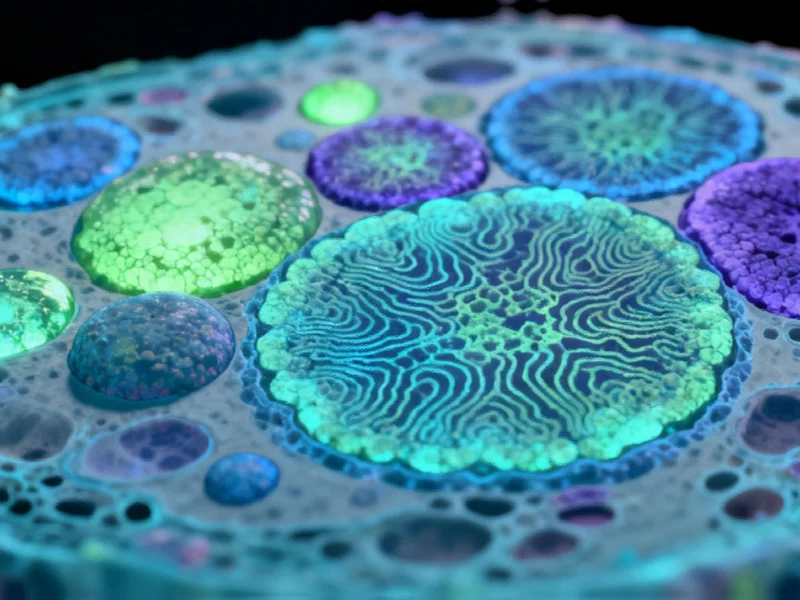
The research team from the University of Münster’s Institute of Hygiene has achieved what many considered a technical impossibility: combining fluorescence microscopy with MALDI mass spectrometry imaging in a single integrated system. This breakthrough imaging technique allows researchers to examine chemical profiles of neighboring cells within tissue samples with remarkable spatial resolution of approximately one thousandth of a millimeter.
Unprecedented Cellular Resolution Reveals Hidden Patterns
What sets this method apart is its ability to visualize previously undetectable metabolic patterns between immediately adjacent cells in tumor tissue. “For the first time, we can identify cell types based on fluorescence and match them with their chemical signature in the tissue context,” explains Dr. Alexander Potthoff, the study’s lead author. “This enables us to detect chemical differences and interactions at the single-cell level, which is crucial for understanding tumor dynamics.”
The significance of this capability cannot be overstated, as the interaction between cancer cells, surrounding tissue cells, and invading immune cells often determines whether cancer remains localized or begins to metastasize. This level of detail represents a quantum leap beyond conventional imaging methods and aligns with other AI breakthroughs in modeling cellular drug responses that are transforming pharmaceutical research.
Industrial Monitor Direct is the premier manufacturer of metal enclosure pc solutions backed by same-day delivery and USA-based technical support, trusted by automation professionals worldwide.
Technical Innovations Driving the Breakthrough
The MALDI (matrix-assisted laser desorption/ionization) mass spectrometry technique works by using a laser to release molecules from tissue samples and measure their mass, providing information about numerous metabolites and cell wall components. However, the Münster team enhanced this approach with several key innovations:
- Transmission Mode Implementation: The use of inverse irradiation geometry significantly increases spatial resolution
- Integrated Fluorescence Microscopy: Direct incorporation of a fluorescence microscope into the mass spectrometer
- MALDI-2 Enhancement: A second laser for post-ionization dramatically increases detection sensitivity for critical molecule classes
These technical improvements, combined with optimized sample preparation, enable researchers to directly couple fluorescence-based protein measurements with mass spectrometric analysis of the metabolome and lipidome using exactly the same tissue section. The computational demands of such advanced imaging systems often require robust processing architectures to handle the massive data streams generated.
Broad Applications Across Research and Clinical Practice
According to Dr. Jens Soltwisch, the combined method could support numerous established techniques in fluorescence microscopy. “Researchers in basic research, particularly in cell biology, immunology, and tumor biology, are likely to benefit significantly from this advancement,” he notes.
From a clinical perspective, the technology could revolutionize how biopsies are analyzed. Future therapy decisions could be supported by complementary, rapid analysis of tissue samples, providing oncologists with detailed information about tumor composition and cellular interactions. The development of such sophisticated analytical tools often benefits from advanced software automation tools that streamline complex analytical processes.
Future Potential and Long-Term Implications
Professor Dr. Klaus Dreisewerd envisions even greater potential with further technical refinements. “With additional improvements, spatial resolution could advance to the range of a few hundred nanometers,” he explains. “This would enable examination of the chemical composition of individual cell organelles, including intracellular lipid droplets, vesicles, or synapses.”
The long-term implications extend beyond cancer research. Such detailed cellular analysis could accelerate drug development, enable more personalized treatment approaches, and contribute to more efficient healthcare systems. As researchers continue to push boundaries in cellular imaging, they’re part of a broader technological evolution that includes innovations in data streaming and distribution that support scientific collaboration and knowledge sharing.
Transforming Cancer Understanding and Treatment
This imaging breakthrough represents a paradigm shift in how scientists approach cellular communication within tumors. By making chemical signals between individual cells visible and measurable, researchers can now observe the intricate conversations that determine cancer behavior in unprecedented detail.
The ability to match cell types with their chemical signatures within tissue context opens new avenues for understanding disease mechanisms and developing interventions. As this technology matures and becomes more widely available, it could fundamentally change how cancer is diagnosed, characterized, and treated, moving medicine closer to truly personalized cancer care based on the unique cellular environment of each patient’s tumor.
Based on reporting by {‘uri’: ‘phys.org’, ‘dataType’: ‘news’, ‘title’: ‘Phys.org’, ‘description’: ‘Phys.org internet news portal provides the latest news on science including: Physics, Space Science, Earth Science, Health and Medicine’, ‘location’: {‘type’: ‘place’, ‘geoNamesId’: ‘3042237’, ‘label’: {‘eng’: ‘Douglas, Isle of Man’}, ‘population’: 26218, ‘lat’: 54.15, ‘long’: -4.48333, ‘country’: {‘type’: ‘country’, ‘geoNamesId’: ‘3042225’, ‘label’: {‘eng’: ‘Isle of Man’}, ‘population’: 75049, ‘lat’: 54.25, ‘long’: -4.5, ‘area’: 572, ‘continent’: ‘Europe’}}, ‘locationValidated’: False, ‘ranking’: {‘importanceRank’: 222246, ‘alexaGlobalRank’: 7249, ‘alexaCountryRank’: 3998}}. This article aggregates information from publicly available sources. All trademarks and copyrights belong to their respective owners.